

Breathe Easily and Freely
Why Working with the Chest Wall Is Key to Whole-Body Health
An author’s perspective on anatomical relationships, functional assessment, and careful practice – Article Written by Larisa Zhilina
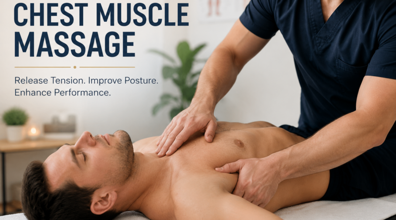
In 2023, I completed the International Massage Teacher Advanced program. My graduation paper was entitled “Working with the Chest Wall.” In this article, I would like to explain why I consider this area one of the most important in massage practice and what changes careful, skillful work may produce.
To me, working with the chest wall is not an isolated technique; it is part of a whole-person approach. Breathing, trunk position, shoulder-girdle movement, abdominal muscle function, and intertissue glide all meet in this region. It should therefore be assessed in connection with the client’s movement, well-being, and reason for seeking treatment.
Anatomical and Physiological Rationale
The thoracic cavity protects vital organs, including the heart, lungs, esophagus, and great vessels. Its osteocartilaginous framework consists of the thoracic spine, ribs, and sternum, while its movement depends on the joints, diaphragm, intercostal and associated muscles, fascia, and skin. The chest wall therefore has both protective and respiratory functions [1,2].
Chest wall position and mobility affect the mechanical environment in which the respiratory muscles and surrounding soft tissues work. When the position of the thorax changes gradually—not necessarily after an injury, but over months or years—the body adapts to a different mechanical strategy. This may be accompanied by persistent functional tension, an altered breathing pattern, changes in shoulder position, and a different distribution of load through the trunk.
In my graduation paper, I emphasized that long-standing changes in this region may become part of a broader pattern of dysfunction. However, the position of the ribs or the density of soft tissues cannot be used to diagnose internal-organ disease or to claim that the chest wall itself has caused pathology. An experienced massage practitioner can identify functional changes, help improve soft-tissue mobility, and recommend medical evaluation when symptoms fall outside the scope of massage practice.
Clinical and Practical Effects of Chest Wall Work
The effects described below were central to my graduation paper and remain important in my practice. Some have a clear anatomical and physiological rationale; others are practical observations. Every client responds differently, so these should be understood as possible effects rather than guaranteed outcomes.
Small randomized studies of manual techniques applied around the diaphragm have reported short-term changes in chest wall and diaphragmatic mobility and in selected functional outcomes [3,4]. A systematic review describes this as a promising area while also emphasizing the heterogeneity of methods and the limited evidence base [5]. These findings support cautious professional interest, but they cannot be generalized to every technique or every client.
Reducing Pelvic Congestion
One less obvious but important effect I consider is the possibility of improving the conditions for venous and lymphatic return from the pelvic region. During breathing, diaphragm movement and changes in intrathoracic and intra-abdominal pressure contribute to the so-called respiratory, or thoracoabdominal, pump. This mechanism supports the movement of venous blood and lymph. Pelvic floor muscle activity is coordinated with breathing and changes in abdominal pressure [2,6,7].
After fascial work with the chest wall, some clients develop freer respiratory excursion and a different pattern of interaction among the myofascial chains of the trunk. This may create more favorable conditions for natural circulation and reduce a subjective sense of tension or “congestion” in the pelvic region.
In practice, I regard this as a supportive approach for clients who report painful menstruation, during preparation for pregnancy, and during postpartum recovery. Chest wall massage is not, however, a stand-alone treatment for gynecological disease, a proven method of eliminating pelvic venous congestion, or a replacement for postpartum rehabilitation. Pain, menstrual changes, pregnancy complications, and new postpartum symptoms require assessment by an appropriately qualified professional.
Supporting Oxygenation and Metabolic Processes
Chest wall work may encourage deeper, calmer, and more efficient breathing. If limited inhalation is partly related to muscular tension, reduced rib mobility, or soft-tissue discomfort, careful manual work may reduce breathing effort and allow freer chest wall excursion.
More complete ventilation creates favorable conditions for oxygen delivery, tissue nutrition, recovery, and normal metabolic reactions. This is the relationship I described in my original paper. At the same time, a healthy person’s resting oxygen saturation is usually already within the normal range. A subjective sense of deeper breathing does not therefore always mean a measurable increase in oxygenation or a faster metabolic rate; objective methods are required to confirm such changes.
Effects on Trunk and Upper-Limb Muscles
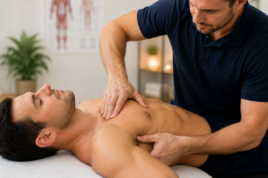
The chest wall provides attachment sites and a region of mechanical interaction for rectus abdominis, the oblique muscles, and transversus abdominis. Through the costal margins, sternum, fascial layers, and intra-abdominal pressure, these muscles contribute to breathing, movement, and trunk stabilization. After fascial work, excessive tension may decrease, tissue glide may improve, and muscular coordination may become more balanced. In practice, this is often described as a return toward normal resting tone, which may have a positive effect on posture and trunk stability.
The shoulder girdle is also functionally connected with the thorax. Pectoralis major and minor, serratus anterior, latissimus dorsi, the intercostal tissues, and the thoracic spine all contribute to scapular position and shoulder movement. Some clients therefore develop greater or more comfortable shoulder rotation after work in this region. I understand this not as an isolated effect on a single rotator but as a change in overall scapulothoracic mechanics and soft-tissue load distribution.
Practical Recommendations
It is useful to begin with a general assessment of breathing, shoulder-girdle position, thoracic mobility, and the client’s response to simple movements. More local tissue assessment can follow. The practitioner should compare findings before and after the intervention and avoid drawing conclusions from a single palpatory sign.
Assessing Tissue Mobility
In fascial work, it is important to assess tissue mobility and identify areas of limited glide between muscles and fascial layers. This can be done by gently shifting one tissue relative to another. If the tissues move with difficulty or feel dense and “stuck,” the finding may suggest local tension, fascial densification, or restricted intertissue glide. It should be recorded as a palpatory observation rather than proof of an adhesion.
In practice, such restrictions may be found near the transition between rectus abdominis and pectoralis major, between the superficial fascia and pectoralis major, and in the region where latissimus dorsi, serratus anterior, the intercostal muscles, the fascial structures of the chest wall, and the thoracolumbar fascia interact.
Assessment should include not only local sensations but also overall chest wall mobility, shoulder-girdle position, and the breathing pattern. A dense area may have a different functional significance in one client than in another.
Functional Testing
Strength and muscular-endurance tests that fall within the practitioner’s training can be useful. They provide a more objective baseline and help show how function changes after manual work. Combining palpation with movement, strength, and endurance tests provides a fuller picture than relying only on perceived tenderness or tissue density.
For a before-and-after comparison, the practitioner may select one or two reproducible movements, such as thoracic rotation, shoulder flexion or abduction, internal or external rotation, together with a subjective rating of breathing ease. Testing should not provoke pain and must remain within the massage practitioner’s professional competence.
A reduction in strength does not always mean that a muscle is “weak” because of a fascial adhesion. Pain, protective tension, altered coordination, joint position, fatigue, and nervous-system state can all influence performance. Test results should therefore be interpreted in context.
Working with the Intercostal Muscles
The intercostal muscles require particular care. Intercostal nerves and blood vessels travel within the chest wall, with the neurovascular bundle close to the inferior border of a rib. Contact should therefore be careful, precisely directed, and appropriately dosed. Excessive pressure, deep focal compression, and aggressive techniques should be avoided, especially when pain, inflammation, injury, or increased sensitivity is present.
Intercostal muscle tension may restrict rib mobility and subjectively make breathing more difficult. Gentle work with the chest wall, ribs, and surrounding muscles can sometimes reduce a sense of tightness and make inhalation and exhalation more comfortable.
Intercostal pain, however, is not always muscular. It may have neurological, articular, inflammatory, or cardiopulmonary causes. If the nature of the pain is uncertain, manual work should stop and medical assessment should be recommended.
Possible Changes After a Session
After chest wall work, a client may report:
- freer and calmer breathing;
- less perceived tightness in the chest;
- improved shoulder-girdle mobility;
- a change in habitual trunk and shoulder position;
- a sense of relaxation in the abdominal and chest wall muscles.
Some changes may be noticeable immediately, although their magnitude depends on the client’s baseline condition, goals, the techniques selected, and individual response. To avoid relying only on an impression, it is useful to repeat the same movement tests and subjective breathing rating used at the beginning of the session.
Safety and Contraindications
Free breathing is important for well-being and quality of life, but chest wall work should form part of an integrated approach. Before the session, the practitioner should take a health history and ask about symptoms, injuries, surgery, medical conditions, medication, and contraindications. Before contacting the anterior or lateral chest, the practitioner should explain the purpose and boundaries of the work, provide professional draping, and obtain the client’s clear consent.
The session should be postponed or discussed with a physician when there is acute infection or fever, recent chest trauma or a suspected rib fracture, active inflammation or a skin lesion in the treatment area, recent surgery, an unstable cardiopulmonary condition, marked osteoporosis, a bleeding disorder, known thrombosis, a complicated pregnancy, or new symptoms in the early postpartum period [8,10].
Stop the massage immediately and seek urgent medical help if new or worsening chest pain or pressure, marked shortness of breath, a cold sweat, nausea, light-headedness, unusual weakness, a rapid or irregular heartbeat, numbness, or pain radiating to an arm, the back, neck, or jaw occurs [9]. Sudden unexplained breathlessness, chest pain that worsens with a deep breath or cough, coughing up blood, or fainting also requires urgent medical evaluation [10].
Conclusion
Chest wall work may influence not one isolated symptom but the relationship among breathing, trunk position, shoulder-girdle movement, and the myofascial chains. This is why I consider the region a key area in massage practice. Its value becomes most apparent when the practitioner carefully assesses the baseline, works gently and precisely, and then checks the client’s functional response.
Stay healthy, and work with care, guided by anatomy, functional assessment, and client safety.
The article is translated into English by ChatGPT
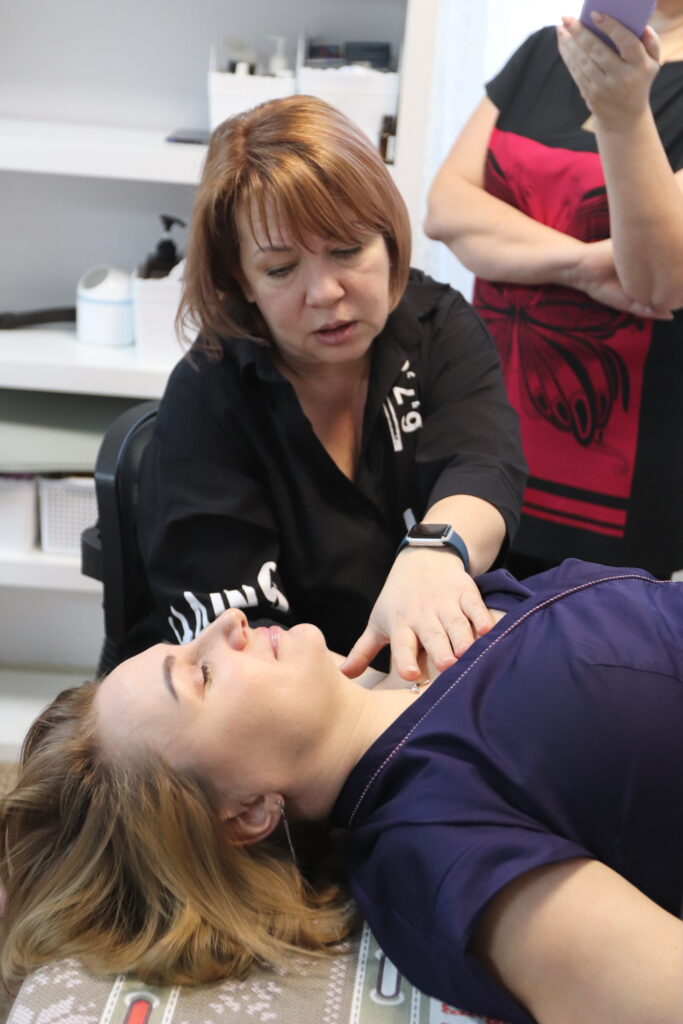
Thank you to Larisa Zhilina for this article.
References
- Donley ER, Holme MR, Loyd JW. Anatomy, Thorax, Wall Movements. In: StatPearls [Internet]. Treasure Island (FL): StatPearls Publishing; 2026. Updated March 13, 2024. https://www.ncbi.nlm.nih.gov/books/NBK526023/
- Bains KNS, Kashyap S, Lappin SL. Anatomy, Thorax: Diaphragm. In: StatPearls [Internet]. Treasure Island (FL): StatPearls Publishing; 2026. Updated July 24, 2023. https://www.ncbi.nlm.nih.gov/books/NBK519558/
- Marizeiro DF, Florêncio ACL, Nunes ACL, Campos NG, de Paula Lima PO. Immediate effects of diaphragmatic myofascial release on the physical and functional outcomes in sedentary women: a randomized placebo-controlled trial. J Bodyw Mov Ther. 2018;22(4):924-929. doi:10.1016/j.jbmt.2017.10.008.
- Rocha T, Souza H, Brandão DC, Rattes C, Ribeiro L, Campos SL, et al. The Manual Diaphragm Release Technique improves diaphragmatic mobility, inspiratory capacity and exercise capacity in people with chronic obstructive pulmonary disease: a randomised trial. J Physiother. 2015;61(4):182-189. doi:10.1016/j.jphys.2015.08.009.
- Fernández-López I, Peña-Otero D, Atín-Arratibel MÁ, Eguillor-Mutiloa M. Effects of Manual Therapy on the Diaphragm in the Musculoskeletal System: A Systematic Review. Arch Phys Med Rehabil. 2021;102(12):2402-2415. doi:10.1016/j.apmr.2021.03.031.
- Hodges PW, Sapsford R, Pengel LH. Postural and respiratory functions of the pelvic floor muscles. Neurourol Urodyn. 2007;26(3):362-371. doi:10.1002/nau.20232.
- Emerich Gordon K, Reed O. The Role of the Pelvic Floor in Respiration: A Multidisciplinary Literature Review. J Voice. 2020;34(2):243-249. doi:10.1016/j.jvoice.2018.09.024.
- National Center for Complementary and Integrative Health. Massage Therapy: What You Need to Know. Accessed August 16, 2026. https://www.nccih.nih.gov/health/massage-therapy-what-you-need-to-know
- American Heart Association. Warning Signs of a Heart Attack. Reviewed December 12, 2024. Accessed August 16, 2026. https://www.heart.org/en/health-topics/heart-attack/warning-signs-of-a-heart-attack
- Centers for Disease Control and Prevention. About Venous Thromboembolism (Blood Clots). Updated May 15, 2024. Accessed August 16, 2026. https://www.cdc.gov/blood-clots/about/index.html
Did you like the article? Or did you like taking part of the World Championship in Massage or one of our classes, then please feel free to give us a nice review on Google.